RED BLOOD CELLS (ERYTHROCYTES):
Mature red blood cells are called erythrocytes.
FACTS:
FACTS:
- They are the most common type of blood cell
- Their primary role is to carry oxygen to the tissues of the body and to carry carbon dioxide away from the body via the circulatory system
- They are flexible and elastic, taking up oxygen in the lungs and then carrying to and releasing it in the tissues while squeezing through tiny capillaries
- They are formed in the bone marrow via a process called erythropoiesis, in response to the hormone erythropoietin, which stimulates their production. It is secreted by the kidneys.
- They are broken down in the spleen
- They have a lifespan of up to 120 days
- They are rich in hemoglobin, which carries iron that can bind oxygen, and hemoglobin is the pigment responsible for the red color of red blood cells, and it is recycled
- The cell membrane is made up of proteins and lipids (fats) and contains a cytoskeleton that provides structure and stability
- They are oval, biconcave disks, which increases their surface for O2/CO2 exchange, and they are not nucleated once they mature (they lose their nucleus)
- About 2.4 million new RBCs are produced every second in the average healthy human adult
- The average human adult has 20-30 trillion RBCs, which composes about 1/4th of the entire cell population of the human body
- Their circulation through the human body takes about 20 seconds
- RBCs have an average diameter of about 6.2-8.2 microns (micrometers) and are about 2.0-2.5 microns (micrometers) thick
- Hemoglobin takes up about 1/3rd of the total RBC volume

Healthy, normal, pink, mature erythrocytes (red blood cells)
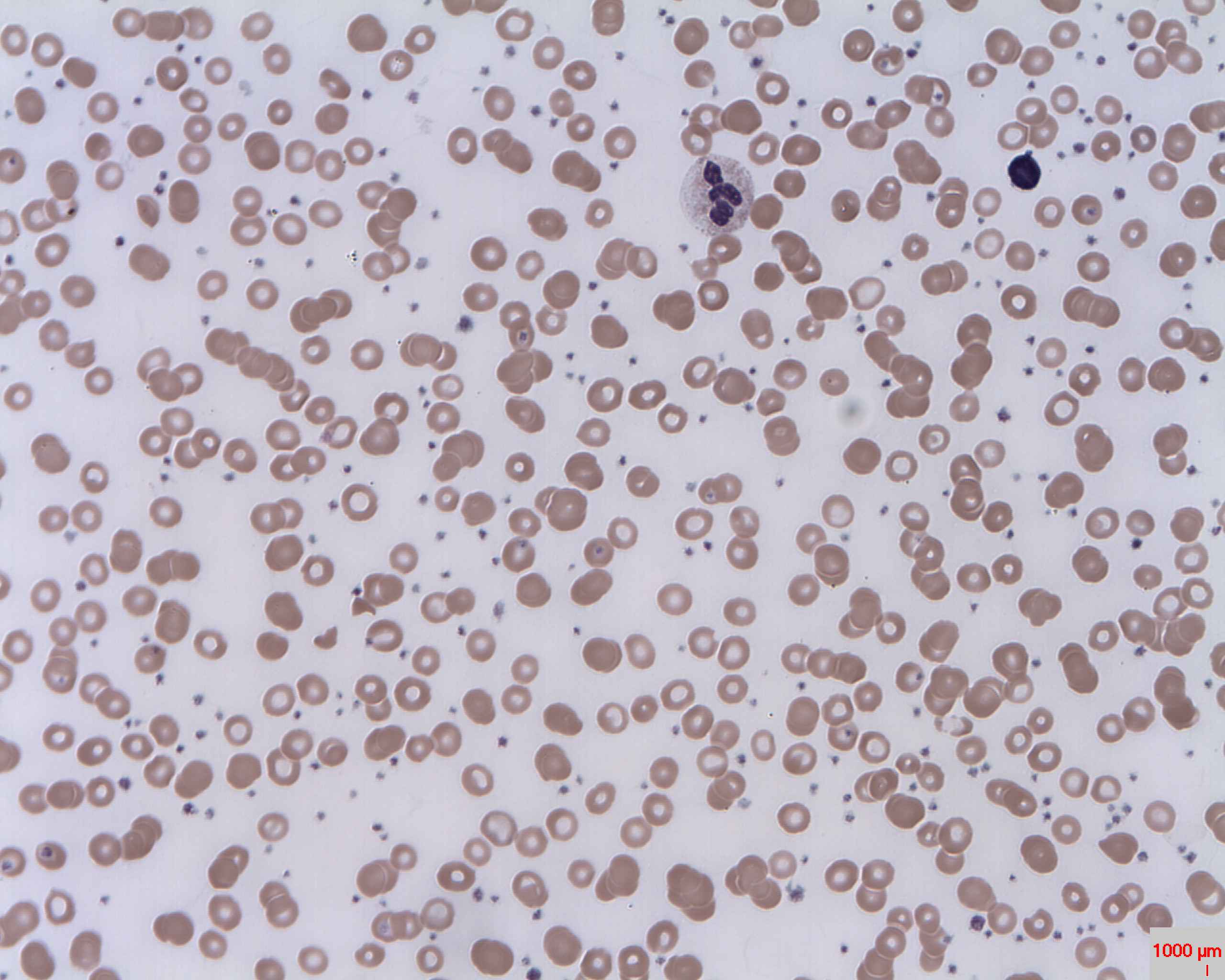
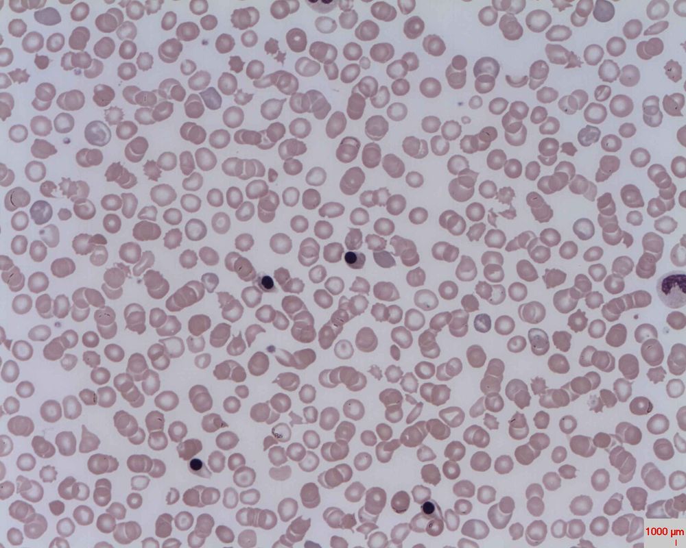
This blood slide shows a variety of red blood cells, some normal, but many abnormal
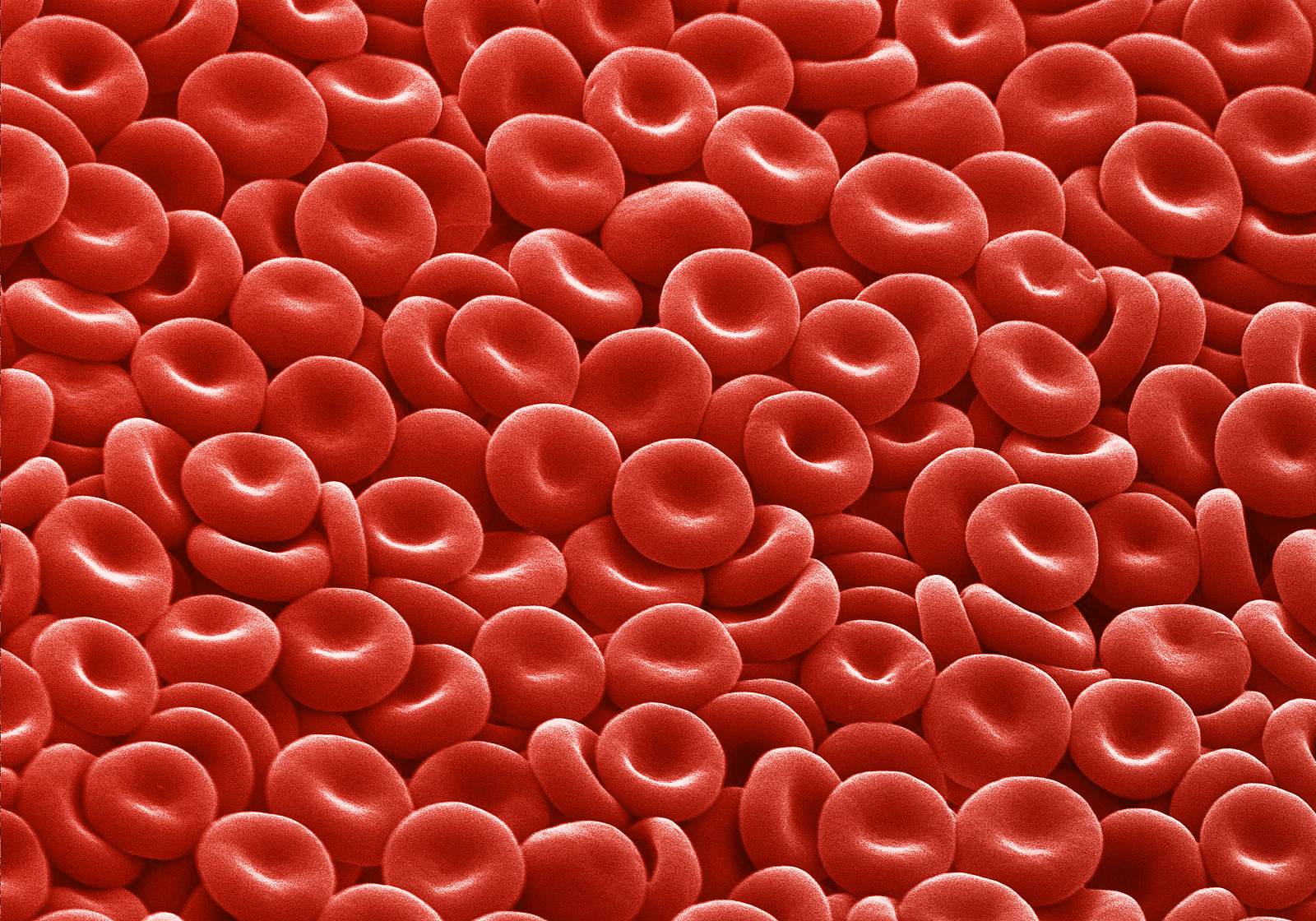
Colorful Scanning Electron Microscope image of red blood cells

Red blood cell membrane

A healthy red blood cell seen by scanning electron microscopy
|

Transmission scanning electron microscope image of red blood cells
|

This is a great image of the discoid shape of the red blood cell. The red color is due to the pigmented hemoglobin molecule.
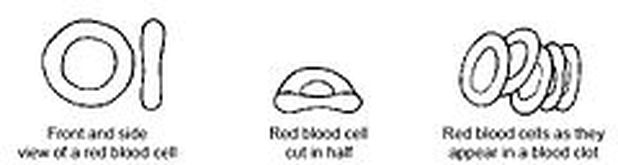
The discoid shape of the red blood cell
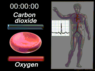
A red blood cell carries oxygen to the cells and tissues of the body and carries carbon dioxide away from the tissues and cells, where it is exhaled by the lungs and excreted by the urinary tract system.
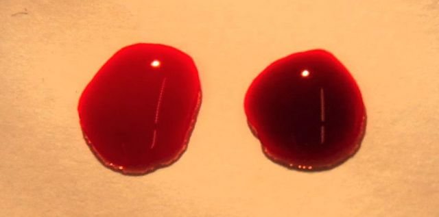
Oxygenated blood (left) and deoxygenated blood (right); The blood you seen in your veins is deoxygenated blood, which appears blue because of the combination of dark red blood and translucent skin
|
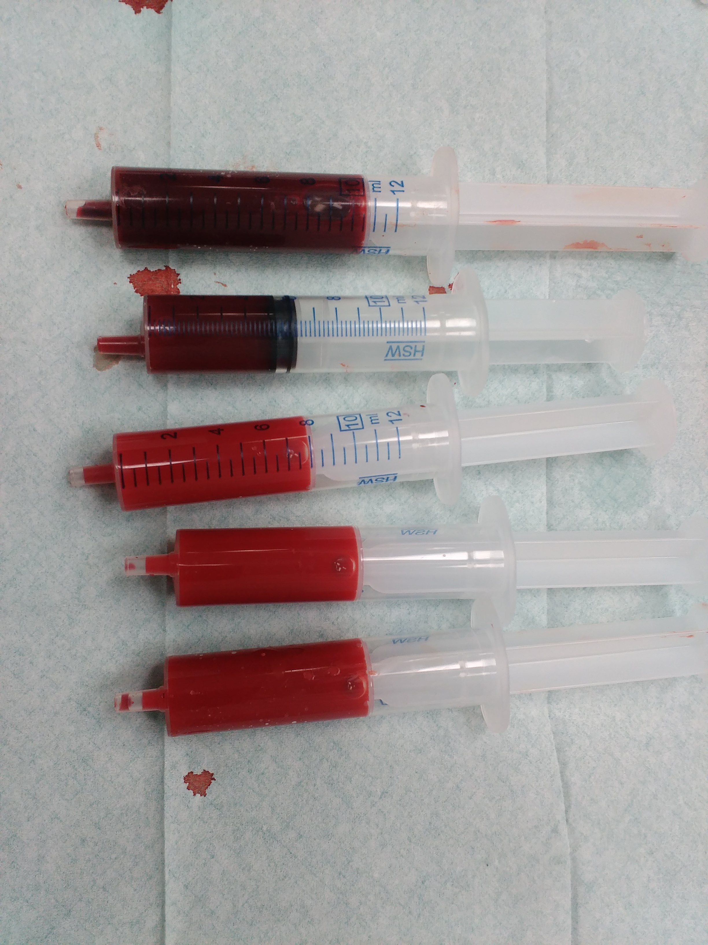
Venous versus arterial blood; Venous blood is darker because it is unoxygenated blood, whereas arterial blood is brighter red because it is oxygenated blood
|
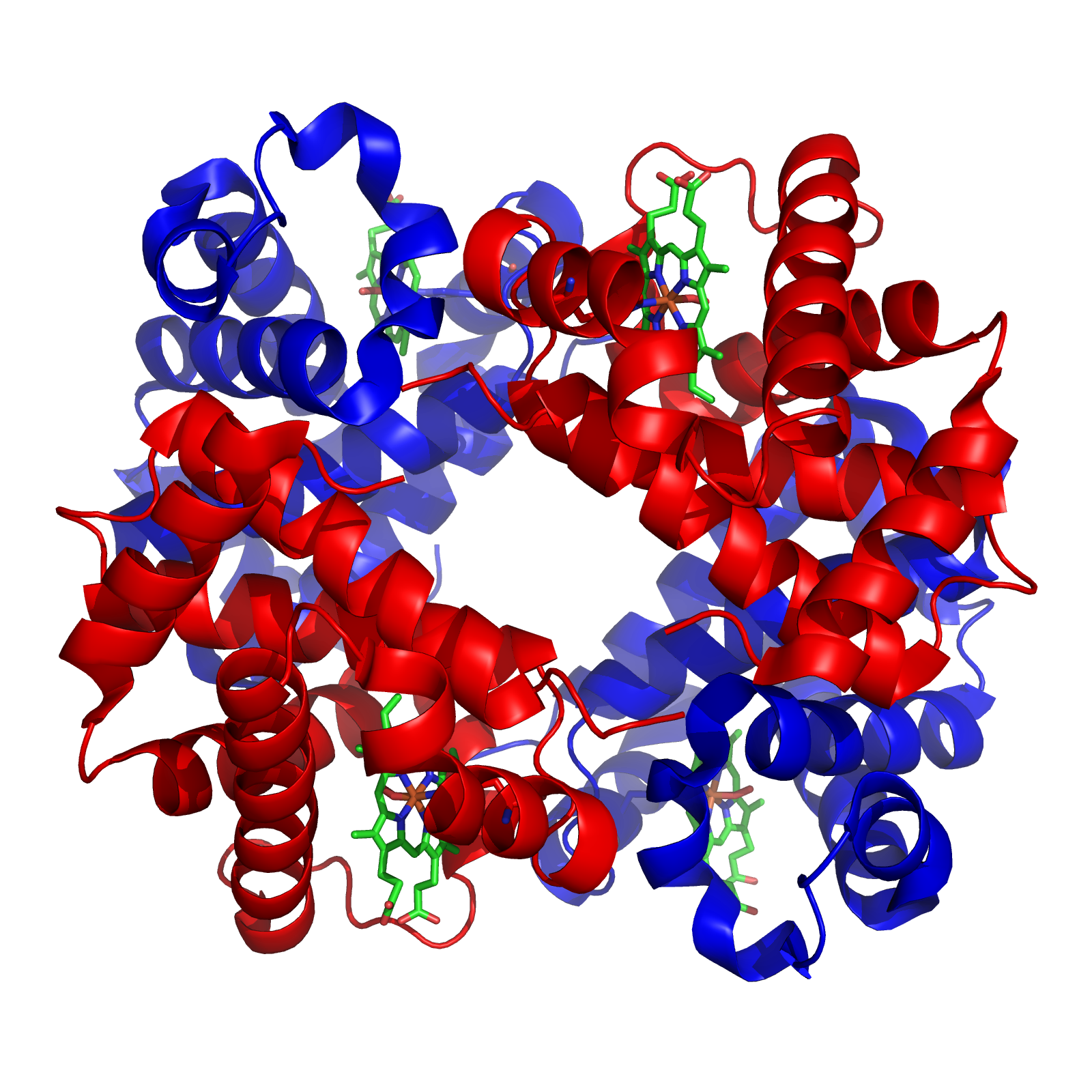
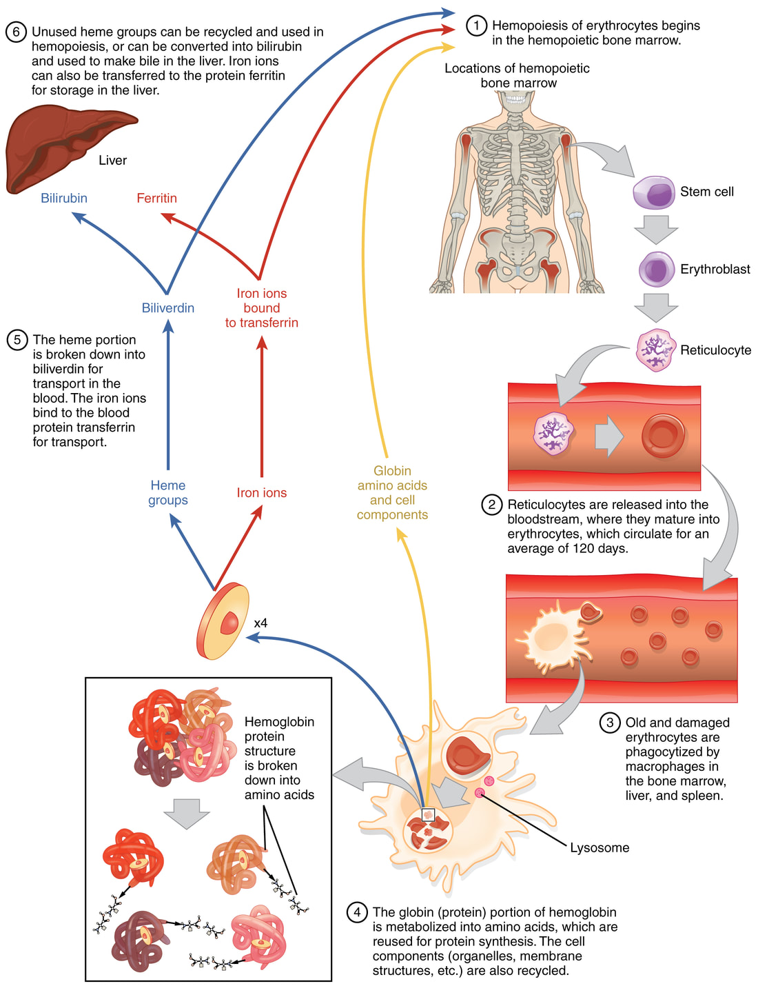
Here is an image of a 4-D hemoglobin molecule; The 4 green chemicals seen within are the 4 iron (Fe) plates that bind oxygen. Each hemoglobin molecule is able to bind 4 oxygen molecules. Red blood cells carry oxygen to the tissues of the body. Red blood cells live about 120 days, then they are destroyed by the spleen, however, the "heme" (iron plates) and "globin" (the 4 chains) are recycled and used again to rebuild new hemoglobin molecules.
An individual with iron-deficiency anemia has either a lower amount of red blood cells than normal, resulting in less hemoglobin, which means less iron and less oxygen, which might be being damaged or destroyed, or they have defective red blood cells, defective hemoglobin molecules, or something has interrupted the iron-oxygen binding capability of the hemoglobin molecules themselves within the red blood cells.
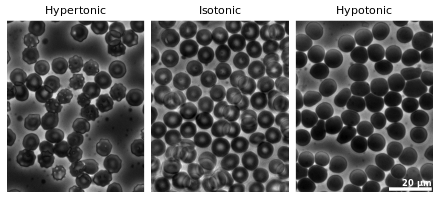
RBC'S AND OSMOSIS:
Red blood cells normally have a nice concave shape, which increases their surface area for carrying oxygen. Red blood cells in an isotonic solution experience no net movement of water in or out of the cell. An isotonic solution is a balanced solution. However, when there is more solute, like sodium chloride (NaCl) outside the cell than inside it, water will rush out of the cell to try to reach an equilibrium (balance), which will cause the cell to shrink or crenate and shrivel up. This is what happens when we get dehydrated. On the other hand, if there is too much solute like NaCl inside the cell, then water will rush into the cell to try to reach equilibrium (balance), and the cell will swell up and may burst (lyse). This could potentially happen if you go to the hospital to be treated for dehydration and they administer IV fluids and don't watch your IV fluid intake carefully. Nurses and assistants check on individuals regularly, and should do so to make sure this does not happen. This is also why there are different types of IV solutions and they are formulated just right to help bring your fluids back into balance. Many of them are a combination of NaCl and glucose (dextrose).
Water moves in and out of the cell by osmosis, the special diffusion of water (the solvent) moving across the semi-permeable and selectively permeable cell membrane. This does not require energy, and is water moving with the concentration gradient. The pressure that generates the process of osmosis is referred to as osmotic pressure.
Water moves in and out of the cell by osmosis, the special diffusion of water (the solvent) moving across the semi-permeable and selectively permeable cell membrane. This does not require energy, and is water moving with the concentration gradient. The pressure that generates the process of osmosis is referred to as osmotic pressure.

Red blood cells in solution
By LadyofHats - did it myself based on [1], [2] ,[3] and [4]., Public Domain, https://commons.wikimedia.org/w/index.php?curid=1685492
Red blood cells in solution, as observed under phase-contrast microscopy; By Zephyris - Own work, CC BY-SA 3.0, https://commons.wikimedia.org/w/index.php?curid=18401754
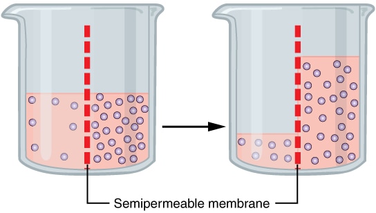
Osmosis: the purple molecules represent the movement of water (solvent) across the semi-permeable membrane to equalize the higher concentration of solute on the other side; By OpenStax - https://cnx.org/contents/[email protected]:fEI3C8Ot@10/Preface, CC BY 4.0, https://commons.wikimedia.org/w/index.php?curid=30131189
ERYTHROPOIESIS:
Erythropoiesis means the production of red blood cells. It is form of hematopoiesis, the production of blood cells. It occurs in response to a hormone called erythropoietin, which is secreted by the adrenal glands which sit on top of the kidneys. It is one of the functions of the kidneys.
PRODUCTION OF RBC'S AND THEIR CYCLE:
- Multipotent hematopoietic stem cell
- These intially develop from the mesoderm in the womb hemangioblast cells
- There are 2 lines that develop from the multipotent HSCs: myeloid and lymphoid
- In adults, these are found in the red bone marrow at the ends of long bones and sternum, and can be found in the peripheral blood and can be found also in the umbilical cord blood and are found to some extent in the thymus gland, liver or spleen
- They are capable of self-renewal and can replenish the blood cells
- They are harvested for stem cell transplants in patients who have certain blood cancers such as leukemia or other blood disorders
- They are undifferentiated
- Myeloid progenitor stem cell
- This is the line that red blood cells develop from
- Limited self-renewal potential
- Endure a few rounds of cell division prior to differentiating into a mature cell, as seen in the image below
- Unipotent stem cell
- Committed at this point to one cell type in the myeloid line
- Pronormoblast
- Also called proerythroblast
- Basophilic normoblast
- This is also called a nucleated red blood cell (NRBC) or a prorubricyte
- This stage is normally only seen in the developing fetus and in newborns
- If seen in a blood smear in an adult, this could indicate that something is wrong and that the demand for production is high in the bone marrow
- Anemia
- Bone marrow cancers (leukemia, lymphoma, myeloma)
- Hypoxemia (lack of oxygen)
- Thalassemia
- Myelofibrosis
- A megaloblast seen at this stage is an unusually large sized erythroblast
- May be linked to a vitamin B12 deficiency (this is called pernicious anemia), deficiency of folic acid (vitamin B9), or both (megaloblastic anemias)
- Polychromatophilic normoblast
- This is also called a rubricyte
- This stage is also only normally seen in the bone marrow
- At this level, there is an increase in the production of hemoglobin
- The cell is still nucleated, but the size of the nucleus starts to shrink
- This is also called a rubricyte
- Orthrochromatic normoblast
- Also called a metarubricyte
- This stage is typically only seen in the bone marrow
- At the end of this stage of development, the nucleus will be extruded from the cell
- Reticulocyte
- This is an immature red blood cell that is larger than a mature erythrocyte, and has a slightly gray-blue tint to it
- Erythrocyte
- This is a mature red blood cell
- This is a mature red blood cell
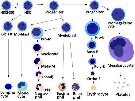
Erythropoiesis

Hematopoiesis; By A. Rad - Own work, CC BY-SA 3.0, https://commons.wikimedia.org/w/index.php?curid=1042490
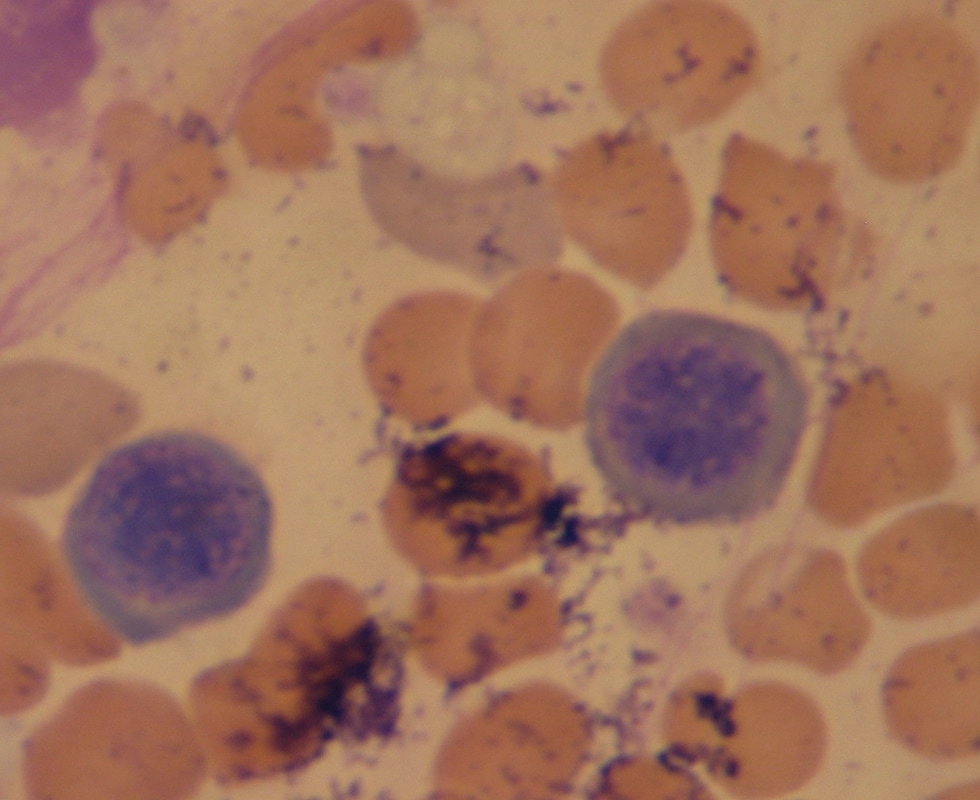
STEM CELL:
PRONORMOBLAST/PROERYTHROBLAST:

|
|
- The cytoplasm stains dark blue
- The nucleus is spherical (round)
- 1-2 nucleoli
- The nuclear chromatin is fine
- The cell size is approximately 12-20 micrometers
- The N/C Ration is 8:1
- Reference:
- Bone Marrow: 1%
- Peripheral Blood: 0% (You should not see these in the peripheral blood. If you do, something is wrong and it could be a type of cancer you need to rule out)
- Anemia
- Myelofibrosis
- Thalassemia
- Tuberculosis
- Bone marrow cancer (leukemia, myeloma, lymphoma)
- Chronic hypoxemia
- Anemia
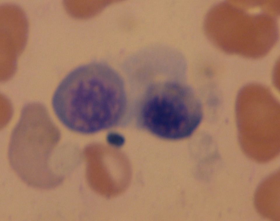
BASOPHILIC NORMOBLAST/BASOPHILIC ERYTHROBLAST:

|
|
- The cytoplasm stains dark blue
- The nucleus is spherical
- There are 0-1 nucleoli
- The chromatin is slightly condensed
- The cell size ranges from 10-15 micrometers
- The N/C Ratio is 6:1
- Reference:
- Bone Marrow: 1-4%
- Peripheral Blood: 0% (You should not see these in the peripheral blood. If you do, something is wrong and it could be a type of cancer you need to rule out)
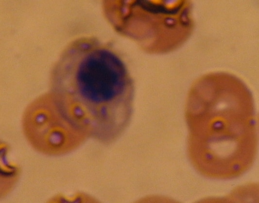
POLYCHROMATOPHILIC NORMOBLAST/POLYCHROMATOPHILIC NORMOBLAST:

|

Nucleated RBC
|
|
- The cytoplasm stains gray-blue
- The nucleus is spherical
- There are 0 nucleoli
- The chromatin is very condensed
- The cell size ranges from 10-12 micrometers
- The N/C ratio is 4:1
- Reference:
- Bone Marrow: 10-20%
- Peripheral Blood: 0% (You should not see these in the peripheral blood. If you do, something is wrong and it could be a type of cancer you need to rule out)
ORTHROCHROMATIC NORMOBLAST:

At this stage, the RBC will extrude its nucleus
|
|
- The cytoplasm stains blue-to-salmon
- The nucleus is spherical
- There are 0 nucleoli
- The chromatin is fully condensed
- The cell size is 8-10 micrometers
- The N/C ratio is 0.5-1.0
- Reference:
- Bone Marrow: 5-10%
- Peripheral Blood: 0% (You should not see these in the peripheral blood. If you do, something is wrong and it could be a type of cancer you need to rule out)
RETICULOCYTE: Polychromatic Erythrocyte

Reticulocytes appear blue in stain. The "reticulum" that is leftover after the RBC extrudes its nucleus at the end of the stage before this one gives it its name. Reticulocytes are slightly larger than the mature erythrocytes.
|

|
- The cytoplasm is Blue-to-salmon in Wright or Wright-Giemsa stain unless stained with Prussian Blue or New Methylene Blue stain as shown above
- The speckles seen in the cells are remnants of the endoplasmic reticulum, hence the name "reticulo"cytes
- The nucleus is absent (*erythrocytes lose their nucleus just prior to this stage)
- There are also no nucleoli
- There is no chromatin
- The cell size is 8.0-8.5 micrometers
- There is no N/C ratio
- Reference:
- Bone Marrow: 1%
- Peripheral Blood: 0.5-2.0%
MATURE ERYTHROCYTE:


- The cytoplasm stains salmon in Wright or Wright-Giemsa stain
- The nucleus is absent
- There are no nucleoli
- There is no chromatin
- The cell size is 7-8 micrometers
- The N/C Ratio is N/A
- Reference:
- Bone Marrow: NA
- Peripheral Blood: Predominant Cell Type
