Overview of Blood:
Blood: It sustains life, it carries iron, it carries oxygen to our brain and tissues of the body, it keeps us healthy and fights off microbes, it clots our blood when we cut or injure ourselves. Blood is crucial to life. We will be discussing what makes blood unique and all the components of normal, healthy blood, as well as abnormalities and how they affect the rest of the body. First, you need to understand the components of blood and what makes up its composition.
Blood carries oxygen, iron and nutrients to the cells and tissues of the body, and it carries CO2 and metabolic wastes such as urea and lactic acid away from the cells and tissues of the body, where it is excreted via exhalation and urine.
Blood consists of red blood cells suspended in a liquid medium called plasma. Plasma contains clotting factors that aid in the coagulation (clotting) of blood. It also contains white blood cells. It accounts for about 7% of the body weight. Each adult has about 5 liters of blood.
Plasma Facts:
The blood pH stays in a very narrow balance of 7.35-7.45. Anything below that is referred to as either metabolic acidosis or respiratory acidosis. Anything above that is referred to as either metabolic alkalosis or respiratory alkalosis.
Blood carries oxygen, iron and nutrients to the cells and tissues of the body, and it carries CO2 and metabolic wastes such as urea and lactic acid away from the cells and tissues of the body, where it is excreted via exhalation and urine.
Blood consists of red blood cells suspended in a liquid medium called plasma. Plasma contains clotting factors that aid in the coagulation (clotting) of blood. It also contains white blood cells. It accounts for about 7% of the body weight. Each adult has about 5 liters of blood.
Plasma Facts:
- 92% WATER (by volume)
- Straw yellow color
- Makes up 55% of blood
- Contains proteins, such as albumin
- The main protein in the blood
- Maintains colloidal osmotic pressure of blood
- Contains glucose
- Contains fatty acids (lipids)
- Contains hormones
- Contains CO2
- Mostly in the form of bicarbonate ion
- Contains minerals in the form of ions
- Electrolytes, such as Ca+, Mg+, K+, Cl-, Ph
- Contains blood cells
- Contains clotting factors
- Contains vitamins
- Contains circulating antibodies
- Regulates the core body temperature
- Makes up 2.7-3.0 liters
The blood pH stays in a very narrow balance of 7.35-7.45. Anything below that is referred to as either metabolic acidosis or respiratory acidosis. Anything above that is referred to as either metabolic alkalosis or respiratory alkalosis.
Components of Blood:
The major (solid) components of blood are:
- Red Blood Cells (a) (erythrocytes)
- Approximately 4.7-6.1 million (male)
- Approximately 4.2-5.4 million (female)
- Carry iron, which binds to oxygen and carries oxygen
- Mature RBCs lack a nucleus and organelles
- Marked by glycoprotein receptors, including those responsible for blood type
- Packed RBCs in fractionated blood are the hematocrit (about 45% of the fractionated blood)
- White Blood Cells (b, c, d) (leukocytes)
- Approximately 4,000-11,000
- Immunity
- Destroy pathogens
- Platelets (the little tiny purple spheres between the red and white blood cells) (thrombocytes)
- 200,000-500,000
- Aid in the clotting or coagulation of blood
- Form a platelet plug
- Covered by a fibrin mesh
- Stop bleeding
- Wall off bacterial invasions

a) Mature erythrocytes (red blood cells), b) polymorphonuclear segmented neutrophil (white blood cell), c) eosinophil (white blood cell), d) basophil (white blood cell); Also seen on the slides are platelets
CENTRIFUGED (FRACTIONATED) BLOOD:
When you go to the doctor and they collect your blood, sometimes they spin down your blood to separate it into 3 different parts or layers that they can test for various things. This is performed by centrifuging your blood (spinning it down) at a high rate of speed (rounds per minute or rpms) in a centrifuge.
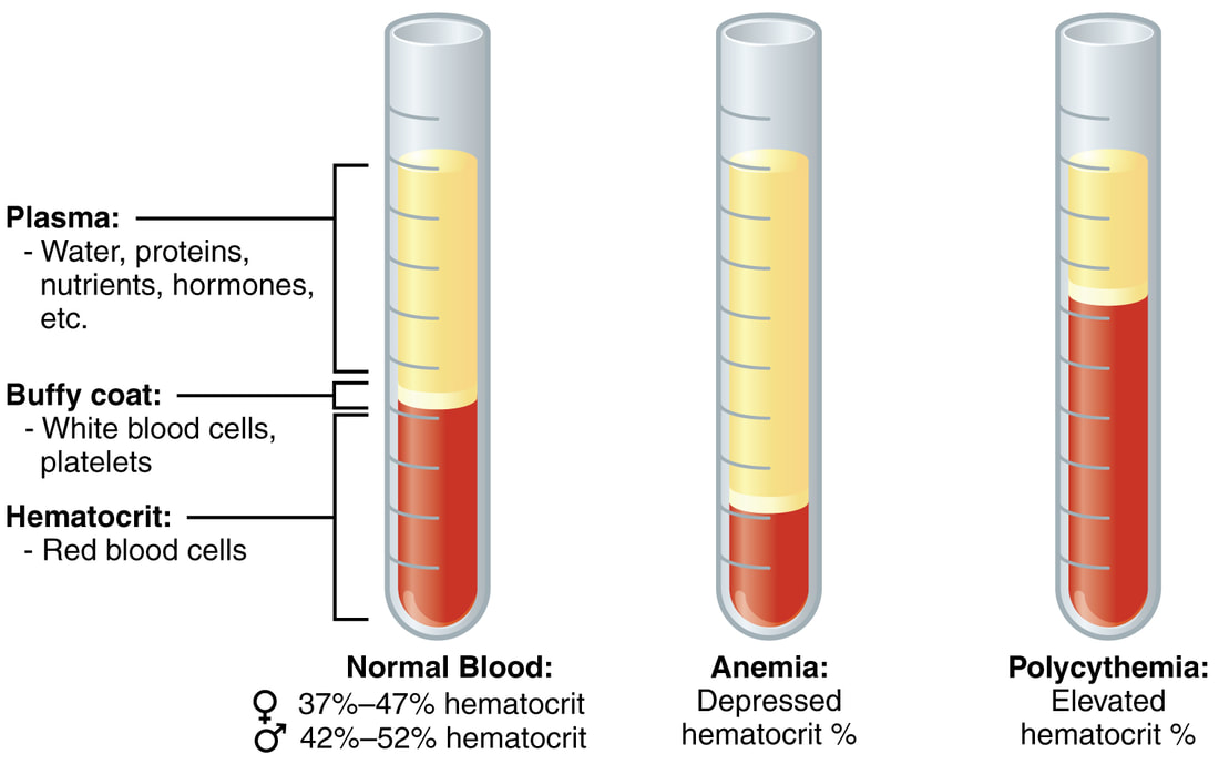
Centrifuged blood divides into three distinct layers:
-It is mostly water (up to 95%)
-It contains dissolved components, including proteins (albumin, fibrinogen, globulin), sodium, glucose, clotting factors, electrolytes, hormones, carbon dioxide
-Because it is intravascular fluid, it helps to maintain osmotic pressure that keeps everything in balance
-This is prepared by spinning or centrifuging the blood in an anticoagulant-coated tube (EDTA, which is the lavender/purple-top tube). This keeps the blood from clotting.
Centrifuged blood divides into three distinct layers:
- Packed red blood cells (bottom/this is referred to as the hematocrit)
- Buffy coat layer (middle/consists of white blood cells, platelets)
- Plasma (straw-colored, fluid portion of blood containing fibrinogen and clotting factors)
-It is mostly water (up to 95%)
-It contains dissolved components, including proteins (albumin, fibrinogen, globulin), sodium, glucose, clotting factors, electrolytes, hormones, carbon dioxide
-Because it is intravascular fluid, it helps to maintain osmotic pressure that keeps everything in balance
-This is prepared by spinning or centrifuging the blood in an anticoagulant-coated tube (EDTA, which is the lavender/purple-top tube). This keeps the blood from clotting.

Blood is collected in Vacutainer tubes. The purple/lavender top Vacutainer tube contains EDTA, an anticoagulant. This prevents the blood from clotting and enables the blood to separate into 3 distinct layers during the centrifugation process.
|

EDTA tube is the purple topped Vacutainer tube
|

SERUM AND SERUM SEPARATOR TUBE (SST):
Serum is the fluid portion of the blood that DOES NOT contain the clotting factors. It contains all the proteins NOT used for coagulation/clotting. It DOES NOT contain the red or white blood cells or platelets. It is basically the blood plasma MINUS the fibrinogens. It is used for many tests in the clinical laboratory.
A serum separator tube (SST), gold or marbled tiger top Vacutainer tube, is used to separate the blood into serum and blood cells. The SST contains gel that moves between to separate the blood cells and serum during the centrifugation process. It also contains clot activator. Centrifugation and clot activator speed up the natural blood clotting process, which would otherwise take about an hour to occur if left on its own. The gel also contains particles that help the blood to clot quickly so that testing on the serum can begin right away, and has an intermediate density between the blood cells and blood plasma. Therefore, during the spinning process, the blood cells sink to the bottom of the tube and the gel moves between the blood cells and serum separating them into two entities. The gel prevents the blood cells and serum from remixing during the transportation process.
Coagulated blood is clotted blood that yields serum without fibrinogen, but some of the clotting factors remain.

A tiger top SST tube with clot activator BEFORE centrifugation (blood just drawn). Notice how the gel starts out at the bottom of the tube before centrifugation.
|

SST II Vacutainer with clot activator gel AFTER centrifugation, separating the blood cells (bottom) from the serum (top). Notice how the gel has moved between the two components to separate them during the centrifugation process.
|
Serum: Icteric, Lipemic and Hemolytic
After centrifugation of blood into its components by a SST (serum separator tube), the serum may appear something other than clear. It is helpful to be able to recognize these differences because sometimes they can interfere with Chemistry tests.
Icterus (Icteric Specimens):
As seen in the images below, icterus or hyperbilirubenemia (increased bilirubin levels) ranges from 1+ to 4+. Normal, straw-colored serum can be seen on the far left (0). As levels of bilirubin are present or increase, the color of the serum ranges from light gold to amber to dark gold to brownish. This can indicate jaundice or a liver problem, which may range from a virus to alcoholism to cirrhosis to a fluke to a gallstone or hemolytic anemia. The following are tests that may be affected by an icteric sample:
- Mg++ (increased)
- Cholesterol (decreased)
- Triglycerides (decreased)
- Creatinine (decreased)
- Bile acids (decreased)
- Lipase (decreased)
- Total protein (decreased)
- Uric acid (decreased)
- GGT (decreased)
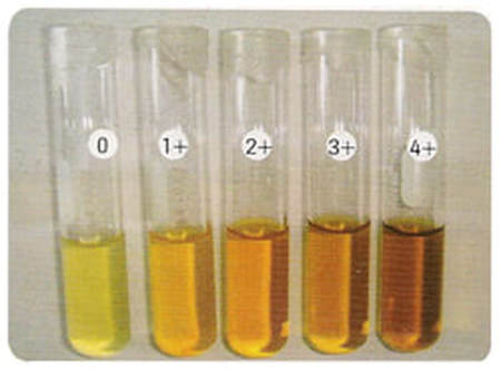
Normal serum (far left) followed by icteric specimens ranging from 1+ to 4+
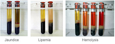
In all specimens, the normal serum is shown on the left, followed by the abnormal serum specimens; 1) Jaundice/Icterus, 2) Lipemia, 3) Hemolysis; http://clinical-laboratory.blogspot.com/2013/06/preventing-pre-analytical-errors.html
Hemolytic Specimens:
Hemolysis is a very common finding in laboratory specimens and is a frequent cause of specimen inadequacy and rejection. Hemolysis is when red blood cells rupture, releasing the hemoglobin pigment, causing the serum to appear pink to orange to red-orange to cherry red. Hemolysis may be intravascular (occur within the patient's veins) or extravascular (outside the veins, in between the cells, or in the specimen itself during centrifugation or mishandling of a specimen or during the phlebotomy blood collection process).
Hemolysis interferes with the spectrophotometric properties of many laboratory instruments and can cause results to be inaccurate. It can actually dilute the serum or plasma. The following tests may be affected by moderate to severe hemolysis:
Decreased Values:
Increased Values:
Lipemic Specimens (Lipemia):
In an individual with increased fats, cholesterol or triglycerides in the bloodstream, the serum or plasma components may appear lipemic, thick and cloudy or milky and turbid due to the presence of these excess fats. Tests that are especially affected include the electrolytes sodium (Na+) and potassium (K+). Hemolysis often occurs along with lipemia because lipemia greatly enhances rupture of red blood cells. The following tests are affected:
Decreased Values:
Increased Values:
Hemolysis is a very common finding in laboratory specimens and is a frequent cause of specimen inadequacy and rejection. Hemolysis is when red blood cells rupture, releasing the hemoglobin pigment, causing the serum to appear pink to orange to red-orange to cherry red. Hemolysis may be intravascular (occur within the patient's veins) or extravascular (outside the veins, in between the cells, or in the specimen itself during centrifugation or mishandling of a specimen or during the phlebotomy blood collection process).
Hemolysis interferes with the spectrophotometric properties of many laboratory instruments and can cause results to be inaccurate. It can actually dilute the serum or plasma. The following tests may be affected by moderate to severe hemolysis:
Decreased Values:
- Troponin
- Bilirubin
- Amylase
- Haptoglobin
- Bicarbonate
Increased Values:
- Potassium (K+)
- Magnesium (Mg+)
- Calcium (Ca++)
- Lactate dehydrogenase (LDH)
- Creatine Kinase (CK)
- AST
- ALT
- Total protein (TP)
- Iron (Fe)
- Phosphate
- Albumin
- Alkaline phosphatase (ALP)
Lipemic Specimens (Lipemia):
In an individual with increased fats, cholesterol or triglycerides in the bloodstream, the serum or plasma components may appear lipemic, thick and cloudy or milky and turbid due to the presence of these excess fats. Tests that are especially affected include the electrolytes sodium (Na+) and potassium (K+). Hemolysis often occurs along with lipemia because lipemia greatly enhances rupture of red blood cells. The following tests are affected:
Decreased Values:
- Sodium (Na+)
- Potassium (K+)
- Chloride (Cl-)
- Bicarbonate
- Lactate dehydrogenase (LDH)
Increased Values:
- Direct bilirubin
- Total bilirubin
- Magnesium (Mg++)
